This Library Guide supports the College of Medicine's curriculum for the Foundational and Clinical Sciences.URL:https://libguides.cmich.edu/med_disciplines
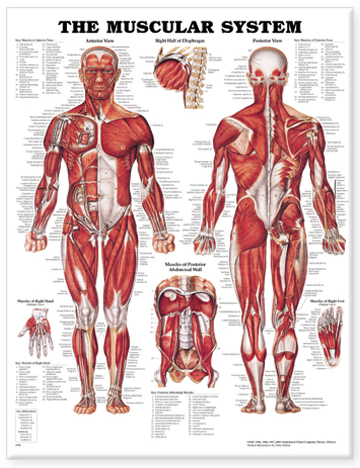
Muscular System Anatomical Chart by Anatomical Chart Co.; Bachin P, ill.
Publication Date: 2000
CMED Learning Commons (Mount Pleasant Campus)
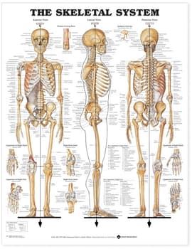
Skeletal System Anatomical Chart by Anatomical Chart Co.; Bachin P, ill.
Publication Date: 2000
CMED Learning Commons (Mount Pleasant Campus)
Recommended Textbooks
The Developing Human: Clinically Oriented Embryology (11e) by Moore KL, Persaud TVN, Torchia MGExtensively revised to incorporate recent research and current clinical practice, The Developing Human: Clinically Oriented Embryology, 11th Edition, covers all aspects of normal and abnormal embryonic and fetal development. In a clear, concise manner and lavishly illustrated throughout, this textbook is designed to successfully meet the needs of medical and health sciences profession students, as well as those in graduate programs. It provides an easy-to-digest, comprehensive review of what can be a complex and challenging subject. BONUS online features such as multiple-choice questions and narrated animations further support student success. Guides readers month by month and stage by stage through embryo and fetal organ and systems development, using full-color photographs of clinical cases, relevant modern medical imaging, and numerous high-quality supportive figures. Includes many new 3D HD color rendered images of embryos and fetuses, as well as 3D reconstructions of whole embryos. Provides clinically-oriented problems for each chapter, with corresponding answers and explanations designed to facilitate discussion and learning. In addition, multiple-choice questions and answers are available online for supportive self-assessment. Features 18 exceptional color animations, now with narration, to help students as they explore and learn the complexity of embryological development. Provides the knowledge base needed for today's examinations, including USMLE Step 1, as well as for future clinical practice. Thoroughly updated information includes new annotations on fundamental molecular events during embryogenesis, now required for many professional examinations. Includes enhanced clinical material in all chapters, with updated tables, Clinical Case highlights and a critical chapter on common signalling pathways during development that covers gene editing (CRISPR/Cas9), induced pluripotent stem cells (iPS), a revision of the sonic hedgehog signalling pathway; and more. Details how molecular biology has affected clinical practice, including techniques such as recumbent DNA technology and stem cell manipulation. Includes an Enhanced eBook version with purchase. Your enhanced eBook allows you to access all of the text, figures, and references from the book on a variety of devices.
Publication Date: 2020
Essential Neuroscience (4e) by Siegel A, Sapru HNEssential Neuroscience integrates must-have neuroscience information with clinical and physiological considerations to help readers master the fundamentals of neuroscience and prepare for board and course exams. Acclaimed for its concise, clinically relevant coverage, this student-friendly book uses a stepwise approach that starts with the basic building blocks of neural anatomy and expands to cover structures and functions, the interaction of systems, and the science of clinical disorders. A well-balanced mix of anatomy, physiology, biology, and biochemistry helps students increase their conceptual understanding of the subject matter and prepare for practice. Vividly illustrated and rich with clinical case studies, summary tables, a glossary of key terms, and comprehensive USMLE-style review questions, this accessible resource fosters the understanding essential to students' success on their exams and in clinical practice. Updated coverage familiarizes you with the latest clinical practices and approaches. Full-color illustrations clarify anatomic structures and complex processes. CT images and MRIs demonstrate radiologic anatomy and present conditions in a clinically relevant context. Clinical Cases enhance your clinical application capabilities and help you confidently manage commonly encountered conditions. Chapter Outlines and Summary Tables emphasize essential content and maximize your study time. Glossary defines bolded key terms at a glance. USMLE-style Review Questions with detailed explanations challenge your understanding and prepare you to excel on course and board exams. eBook available for purchase. Fast, smart, and convenient, today's eBooks can transform learning. These interactive, fully searchable tools offer 24/7 access on multiple devices, the ability to highlight and share notes, and more
Publication Date: 2019
Moore's Clinically Oriented Anatomy (9e) by Dalley II AF, Agur AMRNow with Lippincott Connect! Learn more HERE. Selected as a Doody's Core Title for 2022 and 2023! Renowned for its comprehensive coverage and engaging, storytelling approach, the bestselling Moore's Clinically Oriented Anatomy, 9th Edition, guides students from initial anatomy and foundational science courses through clinical training and practice. A popular resource for a variety of programs, this proven text serves as a complete reference, emphasizing anatomy that is important in physical diagnosis for primary care, interpretation of diagnostic imaging, and understanding the anatomical basis of emergency medicine and general surgery. The 9th Edition reflects the latest changes in the clinical application of anatomy as well as preparation for the USMLE while maintaining the highest standards for scientific and clinical accuracy. NEW! Sex and gender content clarifies important gender considerations and reflects an equitable focus on female as well as male anatomy. Updated medical imaging and integrated surface anatomy within each chapter clearly demonstrates the relationship between anatomy, physical examination, and diagnosis. Extensively revised Clinical Blue Boxes highlight the practical applications of anatomy, accompanied by helpful icons, illustrations, and images that distinguish the type of clinical information covered. Updated introduction establishes the foundational understanding of systemic information and basic concepts essential to success from the classroom to the dissection lab. Revised comprehensive surface anatomy photographs ensure accurate, effective physical examination diagnoses with integrated natural views of unobstructed surface anatomy and illustrations superimposing anatomical structures with landmarks for more accurate physical examination. Insightfully rendered, anatomically accurate illustrations, combined with many photographs and medical images, strengthen comprehension of anatomical concepts and retention of "mental images" of anatomical structures. Bottom Line boxes provide detailed summaries at a glance and underscore the "big-picture" perspective. Illustrated tables clarify complex information about muscles, veins, arteries, nerves, and other structures for easy study and review. Chapter outlines help students find key information quickly and efficiently. Additional online resources: NEW! 20 BioDigital 3D tours provide an ideal self-study experience, allowing students to peel away layers, rotate models, zoom, fade, and test their knowledge with 3D quizzes. More than 100 interactive clinical vignette multiple-choice questions and answers provide valuable self-test opportunities for exam review. Over 140 case studies with questions and explanations engage students in the clinical application process of anatomic principles.
Publication Date: 2023
Netter Atlas of Human Anatomy: Classic Regional Approach (8e) by Netter FHFor clinical professionals or students who are learning anatomy, participating in a dissection lab, sharing anatomy knowledge with patients, or refreshing their anatomy knowledge, the Netter Atlas of Human Anatomy illustrates the body, region by region, in clear, brilliant detail from a clinician's perspective. Unique among anatomy atlases, it contains illustrations that emphasize anatomic relationships that are most important to the clinician in training and practice. Illustrated by clinicians, for clinicians, it contains more than 550 exquisite plates plus dozens of carefully selected radiologic images for common views. This Professional hardcover edition includes access to a downloadable imagebank for personal use. Presents world-renowned, superbly clear views of the human body from a clinical perspective, with paintings by Dr. Frank Netter as well as Dr. Carlos A. G. Machado, one of today's foremost medical illustrators. Content guided by expert anatomists and educators: R. Shane Tubbs, Paul E. Neumann, Jennifer K. Brueckner-Collins, Martha Johnson Gdowski, Virginia T. Lyons, Peter J. Ward, Todd M. Hoagland, Brion Benninger, and an international Advisory Board. Offers region-by-region coverage, including muscle table appendices at the end of each section and quick reference notes on structures with high clinical significance in common clinical scenarios. Contains new illustrations by Dr. Machado including clinically important areas such as the pelvic cavity, temporal and infratemporal fossae, nasal turbinates, and more. Features new nerve tables devoted to the cranial nerves and the nerves of the cervical, brachial, and lumbosacral plexuses. Uses updated terminology based on the second edition of the international anatomic standard, Terminologia Anatomica, and includes common clinically used eponyms. Includes image bank license of full set of Plates and bonus plates from prior editions which allows presentation sharing in groups of 10 or fewer as well as use in scientific poster presentations by active students. Complete Registered User License and institutional licensing information available at NetterReference.com. Also available, alternative versions of the 8th Edition: . Netter Atlas of Human Anatomy: A Systems Approach-paperback+eBook. Same content as the classic regional approach, but organized by organ systems. . Netter Atlas of Human Anatomy: Classic Regional Approach-paperback + eBook. . Netter Atlas of Human Anatomy: Classic Regional Approach with Latin terminology-paperback +eBook. All options contain the same table information and same 550+ illustrated plates painted by clinician artists, Frank H. Netter, MD, and Carlos Machado, MD.
Publication Date: 2022
Wheater's Functional Histology (7e) by O'Dowd G, Bell S, Wright SNow in its seventh edition, Wheater's Functional Histology is designed to give students a working knowledge of histology, including the basic histological structures, tissue types and organ systems. You will learn to recognise the microscopic structure of normal human tissues and understand how this relates to function. The book shows you how to apply histology in a clinical context through coverage of common clinical conditions in each chapter. This must-have book is written clearly and concisely, designed for those studying histology for the first time. Packed with a wealth of illustrations and learning aids, it is a must-have for every medical and health science student. Concise and easy to digest text - easy to follow for those new to histology More than 900 high quality histology images and illustrations, plus detailed explanations to support understanding End of chapter review tables summarising important points Self-assessment questions and rationales to support exam preparation Appendix outlining the basics of microscopy and histological staining techniques Basic glossary An enhanced eBook version is included with purchase. The eBook allows you to access all the text, figures and references, with the ability to search, customize your content, make notes and highlights, and have content read aloud More clinical correlation boxes More clinical images More questions added
Publication Date: 2023
Additional eBooks
The Big Picture: Gross Anatomy, Medical Course & Step 1 Review (2e) by Morton DA, Foreman KB, Albertine KHThe acclaimed book students need to get the BIG PICTURE of Gross Anatomy in the context of healthcare - streamlined and enhanced by online video Gross Anatomy: The Big Picture, Second Edition is the perfect bridge between review and textbooks. With an emphasis on what students truly need to know versus "what's nice to know," it features 450 full-color illustrations that give them a complete, yet concise, overview of essential anatomy. The Second edition is distinguished by the addition of online videos ranging from 3-9 minutes long, and more concise, bulleted text. The book's user-friendly presentation consists of text on the left-hand page and beautiful full-color illustrations on the right-hand page. In this way, you get a "big picture" of anatomy principles, delivered one concept at a time -- making them easier to understand and retain. Striking the perfect balance between illustrations and text, Gross Anatomy: The Big Picture, Second Edition features: *High-yield review questions and answers at the end of each chapter *Numerous summary tables and figures that encapsulate important information *450 labeled and explained full-color illustrations *A final exam featuring 100 Q&As *Important clinically-relevant concepts called to your attention by convenient icons *Bullets and numbering that break complex concepts down to easy-to-remember points
Publication Date: 2019
Essentials of Anatomy and Physiology (8e) by Scanlon VC, Sanders TFlexible presentation of content makes the text suitable for any course. Precisely detailed, full-color illustrations throughout make every concept clear. Highly detailed anatomy photographs and full-color illustrations enhance visual learning. Clinical application boxes put theory into action. Extensive microbiology tables reinforce the text. Questions for every figure in the book encourage critical thinking and reinforce understanding of the image and the content. A wealth of pedagogical features promotes learning and includes chapter outlines * student objectives * new and related clinical terminology (with pronunciations) * study outlines * review questions and "For Further Thought" discussion topics * and a comprehensive glossary. Pretests and post-tests for every chapter with print and email capabilities assess progress.
Publication Date: 2019
Grant's Atlas of Anatomy (16e) by Agur AMR, Dalley II AFIllustrations drawn from real specimens, presented in surface-to-deep dissection sequence, set Grant's Atlas of Anatomy apart as the most accurate illustrated reference available for learning human anatomy and referencing in dissection lab. A recent edition featured re-colorization of the original Grant's Atlas images from high-resolution scans, also adding a new level of organ luminosity and tissue transparency. The dissection illustrations are supported by descriptive text legends with clinical insights, summary tables, orientation and schematic drawings, and medical imaging. Renowned, high-resolution, dynamically colored illustrations organized in dissection sequence enable the formation of 3D constructs for each body region and provide detailed, realistic reference during dissection. Tables detail muscles, vessels, and other anatomic information in an easy-to-use format ideal for review and study. Enhanced medical imaging includes more than 100 clinically significant MRIs, CT images, ultrasound scans, and corresponding orientation drawings to help students confidently apply the laboratory experience to clinical rotations. Color schematic illustrations reinforce the relationships of structures and anatomical concepts in vibrant detail.
Publication Date: 2025
Gray's Anatomy: The Anatomical Basis of Clinical Practice (42e) by Standring SSusan Standring, MBE, PhD, DSc, FKC, Hon FAS, Hon FRCS Trust Gray's. Building on over 160 years of anatomical excellence In 1858, Drs Henry Gray and Henry Vandyke Carter created a book for their surgical colleagues that established an enduring standard among anatomical texts. After more than 160 years of continuous publication, Gray's Anatomy remains the definitive, comprehensive reference on the subject, offering ready access to the information you need to ensure safe, effective practice.? This 42nd edition has been meticulously revised and updated throughout, reflecting the very latest understanding of clinical anatomy from the world's leading clinicians and biomedical scientists. The book's acclaimed, lavish art programme and clear text has been further enhanced, while major advances in imaging techniques and the new insights they bring are fully captured in state of the art X-ray, CT, MR and ultrasonic images. The accompanying eBook version is richly enhanced with additional content and media, covering all the body regions, cell biology, development and embryogenesis - and now includes two new systems-orientated chapters. This combines to unlock a whole new level of related information and interactivity, in keeping with the spirit of innovation that has characterised Gray's Anatomy since its inception. ? Each chapter has been edited by international leaders in their field, ensuring access to the very latest evidence-based information on topics Over 150 new radiology images, offering the very latest X-ray, multiplanar CT and MR perspectives, including state-of-the-art cinematic rendering? The downloadable Expert Consult eBook version included with your (print) purchase allows you to easily search all of the text, figures, references and videos from the book on a variety of devices? Electronic enhancements include additional text, tables, illustrations, labelled imaging and videos, as well as 21 specially commissioned 'Commentaries' on new and emerging topics related to anatomy? Now featuring two extensive electronic chapters providing full coverage of the peripheral nervous system and the vascular and lymphatic systems.? The result is a more complete, practical and engaging resource than ever before, which will prove invaluable to all clinicians who require an accurate, in-depth knowledge of anatomy.
Publication Date: 2021
Gray's Anatomy for Students (5e) by Drake RL, Vogl AW, Mitchell AWM (Eds)Concise, readable text and an outstanding art program make Gray's Anatomy for Students, 5th Edition, your go-to text for essential information in human anatomy. This fully revised volume focuses on the core information medical students need to know, in an easy-access format and with additional multimedia content to facilitate effective study and mastery of the material. A team of expert authors share a wealth of diverse teaching and clinical experience-all enhanced by more than 1,000 innovative, original illustrations by renowned illustrators Richard Tibbitts and Paul Richardson, who capture anatomical features with unrivalled clarity. Helps you understand the practical applications of anatomical concepts through unique coverage of surface anatomy, correlative diagnostic images, and clinical case studies. Contains increased representation of diverse population groups throughout, incorporating a wider range of skin tones and important clinical considerations related to transgender and intersex individuals. Presents anatomy logically by body region - as well as bonus updated eBook chapters for each major body system to facilitate learning from a different perspective. Includes new and improved online materials such as self-assessment questions, medical and physical therapy clinical cases, a unique Interactive Surface Anatomy tool, and more. Provides fully revised and updated clinical content including numerous new In the Clinic boxes, images, and correlates throughout that reflect the latest advances seen in clinical practice. New and updated Clinical Cases are included in the accompanying enhanced eBook. Features an updated neuroanatomy eBook chapter, so you can learn key aspects of this challenging topic in the context of general anatomy. Improves comprehension of complex cranial nerves with a visual map summarizing cranial nerve distribution and function. Offers schematic drawings for key structures and topics in every chapter, providing an additional, simplified approach to introduce each topic-ideal for quick initial understanding and as a guide for your own anatomy drawings. Enables you to quickly review the basic concepts from each chapter with Conceptual Overviews. An eBook version is included with purchase. The eBook allows you to access all of the text, figures and references, with the ability to search, customize your content, make notes and highlights, and have content read aloud. Evolve Instructor site with a downloadable image bank is available to instructors through their Elsevier sales rep or via request at https://evolve.elsevier.com.
Publication Date: 2024
Lippincott Atlas of Anatomy (2e) by Gest TRAlso available in print: QM25 .T36 2020
Featuring more than 800 high-quality, modern images, Lippincott® Atlas of Anatomy is a vibrantly colored regional atlas of human anatomy offering an unprecedented combination of visual aesthetic appeal and anatomical accuracy. A unique art style featuring bright colors, color coding of anatomical elements, judicious use of labeling, and no extraneous text make this book an ideal tool for students. Each plate teaches specific structures and relationships--displayed using artistic techniques such as 'ghosting,' layering, and color coding--providing clear focus on important structures and resulting in easier and faster comprehension. This unique pedagogy, in combination with the beautiful art style, make this anatomy atlas the preferred choice for teaching and learning. This updated new edition enhances students' understanding of anatomical structures and relationships with many new and modified illustrations, new muscle tables, and convenient online access to tables summarizing key information for multiple anatomical structures. Packaged with the atlas is an interactive eBook version with all of the images, including fully searchable legends and labels and 'zoom and compare' features, as well as online labeling exercises that reinforce students' understanding of newly learned concepts and structures. More than 800 color illustrations--including 16 new and over 300 revised--reflect a variety of techniques to give students an unparalleled understanding of the most clinically relevant anatomical relationships. New muscle tables clarify attachments, innervation and more. A systems overview with summary illustrations helps you confidently guide students from surface anatomy and superficial features to deeper structures that support regional dissection sequences. More than 30 clinical images--including x-rays and CT scans--enhance students' clinical readiness with radiographic views of anatomy. An interactive eBook simplifies review with instant access to all images with fully searchable legends and labels, zoom and compare features, and anatomy tables covering muscles, topographic anatomy, bones, joints, arteries, veins, lymphatics, nerves, fascia, and clinical terms for each chapter. Online 'test yourself' labeling exercises on selected plates allow students to test themselves on newly learned concepts and structures. eBook available for purchase. Fast, smart, and convenient, today's eBooks can transform learning. These interactive, fully searchable tools offer 24/7 access on multiple devices, the ability to highlight and share notes, and more
Publication Date: 2020
Netter's Clinical Anatomy (5e) by Hansen JTOffering a targeted, concise approach to clinical anatomy, Netter's Clinical Anatomy, 5th Edition, is a portable, easy-to-read text that bridges normal anatomy to common clinical conditions. It features nearly 600 superb illustrations by Drs. Frank H. Netter and Carlos Machado that provide essential descriptions of anatomy, embryology, and pathology to help you understand their clinical relevance. Authored by John Hansen, PhD, an Honored Member of the American Association of Clinical Anatomists, this visually stunning text covers and reviews the sizable area of human anatomy and pathology in a quick, easy-to-grasp format. Nearly 600 Netter and Machado illustrations provide essential depictions of anatomy, embryology, and pathology to help you quickly learn and understand through visuals. Every clinical correlation is illustrated-more than 200 in all. Clinical Focus boxes present hundreds of illustrated clinical correlations that bridge anatomy to pathophysiology, and Features and Characteristics boxes explain the relation between structure and function. Muscle/Ligament/Joint tables summarize attachment points, actions, and other key information related to each structure. Uses updated terminology based on the second edition of the international anatomic standard, Terminologia Anatomica, and content is aligned with Netter Atlas of Human Anatomy, 8th Edition. Both USMLE-style review questions and short answer questions online help you gauge your mastery of the material and assess areas in need of further study. Other study tools include bonus clinical focus boxes, and 3D models. Enhanced eBook version included with purchase. Your enhanced eBook allows you to access all of the text, figures, and references from the book on a variety of devices. Evolve Instructor site with an image bank of clinical correlates is available to instructors through their Elsevier sales rep or via request at https://evolve.elsevier.com.
Publication Date: 2023
Netter Atlas of Human Anatomy: Classic Regional Approach (8e) by Netter FHFor clinical professionals or students who are learning anatomy, participating in a dissection lab, sharing anatomy knowledge with patients, or refreshing their anatomy knowledge, the Netter Atlas of Human Anatomy illustrates the body, region by region, in clear, brilliant detail from a clinician's perspective. Unique among anatomy atlases, it contains illustrations that emphasize anatomic relationships that are most important to the clinician in training and practice. Illustrated by clinicians, for clinicians, it contains more than 550 exquisite plates plus dozens of carefully selected radiologic images for common views. This Professional hardcover edition includes access to a downloadable imagebank for personal use. Presents world-renowned, superbly clear views of the human body from a clinical perspective, with paintings by Dr. Frank Netter as well as Dr. Carlos A. G. Machado, one of today's foremost medical illustrators. Content guided by expert anatomists and educators: R. Shane Tubbs, Paul E. Neumann, Jennifer K. Brueckner-Collins, Martha Johnson Gdowski, Virginia T. Lyons, Peter J. Ward, Todd M. Hoagland, Brion Benninger, and an international Advisory Board. Offers region-by-region coverage, including muscle table appendices at the end of each section and quick reference notes on structures with high clinical significance in common clinical scenarios. Contains new illustrations by Dr. Machado including clinically important areas such as the pelvic cavity, temporal and infratemporal fossae, nasal turbinates, and more. Features new nerve tables devoted to the cranial nerves and the nerves of the cervical, brachial, and lumbosacral plexuses. Uses updated terminology based on the second edition of the international anatomic standard, Terminologia Anatomica, and includes common clinically used eponyms. Includes image bank license of full set of Plates and bonus plates from prior editions which allows presentation sharing in groups of 10 or fewer as well as use in scientific poster presentations by active students. Complete Registered User License and institutional licensing information available at NetterReference.com. Also available, alternative versions of the 8th Edition: . Netter Atlas of Human Anatomy: A Systems Approach-paperback+eBook. Same content as the classic regional approach, but organized by organ systems. . Netter Atlas of Human Anatomy: Classic Regional Approach-paperback + eBook. . Netter Atlas of Human Anatomy: Classic Regional Approach with Latin terminology-paperback +eBook. All options contain the same table information and same 550+ illustrated plates painted by clinician artists, Frank H. Netter, MD, and Carlos Machado, MD.
Weir and Abrahams' Imaging Atlas of Human Anatomy (6e) by Spratt JD, Salkowski LR, Loukas M, Turmezei T, Weir J, Abrahams PHThe perfect up-to-date imaging guide for a complete and 3-dimensional understanding of applied human anatomy Imaging is ever more integral to anatomy education and throughout modern medicine. Building on the success of previous editions, this fully revised sixth edition provides a superb foundation for understanding applied human anatomy, offering a complete view of the structures and relationships within the whole body, using the very latest imaging techniques. All relevant imaging modalities are included, from plain radiographs to more advanced imaging of ultrasound, CT, MRI, functional imaging and angiography. Coverage is further enhanced by a carefully selected range of BONUS electronic content, including clinical photos and cases, ultrasound videos, labelled radiograph 'slidelines', cross-sectional imaging stacks and test-yourself materials. Uniquely, key syllabus image sets are now highlighted throughout to aid efficient study, as well as the most common, clinically important anatomical variants that you should be aware of. This superb package is ideally suited to the needs of medical students, as well as radiologists, radiographers and surgeons in training. It will also prove invaluable to the range of other students and professionals who require a clear, accurate, view of anatomy in current practice. Fully revised legends and labels and new high-quality images-featuring the latest imaging techniques and modalities as seen in clinical practice Covers the full variety of relevant modern imaging-including cross-sectional views in CT and MRI, angiography, ultrasound, fetal anatomy, plain film anatomy, nuclear medicine imaging and more - with better resolution to ensure the clearest anatomical views Core syllabus image sets now highlighted throughout-to help you focus on the most essential areas to excel on your course and in examinations Unique summaries of the most common, clinically important anatomical variants for each body region-reflects the fact that around 20% of human bodies have at least one clinically significant variant New orientation drawings-to help you understand the different views and the 3D anatomy of 2D images, as well as the conventions between cross-sectional modalities Ideal as a stand-alone resource or in conjunction with Abrahams' and McMinn's Clinical Atlas of Human Anatomy-where new links help put imaging in the context of the dissection room Now a more complete learning package than ever before, with superb BONUS electronic enhancements embedded within the accompanying eBook, including: Labelled image 'stacks'-that allow you to review cross-sectional imaging as if using an imaging workstation Labelled image 'slidelines'-showing features in a full range of body radiographs to enhance understanding of anatomy in this essential modality Self-test image 'slideshows' with multi-tier labelling-to aid learning and cater for beginner to more advanced experience levels Labelled ultrasound videos-bring images to life, reflecting this increasingly clinically practiced technique Questions and answers accompany each chapter-to test your understanding and aid exam preparation 34 pathology tutorials-based around nine key concepts and illustrated with hundreds of additional pathology images, to further develop your memory of anatomical structures and lead you through the essential relationships between normal and abnormal anatomy High-yield USMLE topics-clinical photos and cases for key topics, linked and highlighted in chapters